Cryopreservation and CO2-independent Culture of 3D Cardiac Progenitors for Space Flight Experiments
Application
New method for developing therapeutic stem cells that could repair damaged heart tissue.
Key Benefits
- Potentially improves the ability to generate cardiomyocytes from hiPSCs compared to current methods.
- It may be used for the treatment and/or repair of diseases that damage the heart tissue.
- Culture improvements could apply to other cell types and diseases (e.g., neuron regeneration).
- Expand the basic understanding of various diseases due to improved culture conditions in the life science industry.
Market Summary
Human pluripotent stem cells (hPSCs) can differentiate into cardiomyocytes, thereby a potential source of therapeutic cells to repair damaged heart muscle and help in drug discovery. Understanding the growth and differentiation of cardiomyocyte from human induced pluripotent stem cells in microgravity environment is a great interest given the potential application of these cells in disease modeling, drug development and regenerative medicine. Recent spaceflight experiments showed that human induced pluripotent stem cell-derived cardiomyocytes (hiPSC-CMs) had increased metabolic features and that cardiovascular progenitor cells cultured on the space station increased the cell’s proliferative potential. Study of hiPSCs on space station requires cultivating 3D cardiac progenitors for approximately 3 weeks inside a cell culture module. Although the use of CO2 in cell culture models is standard on Earth supplying CO2 to the cell cultures is difficult on space craft. Carrying CO2 tanks or CO2 incubator on a spaceflight is requires extra space and load capacity. Cell cultures also need to be able to withstand high g-force (up to 3 g) during the launch. In addition, any reduction in extra chores for the astronauts in maintaining experimental setup would be a huge benefit. Thus, there is a need to study hiPSCs without CO2 with minimal setup required.
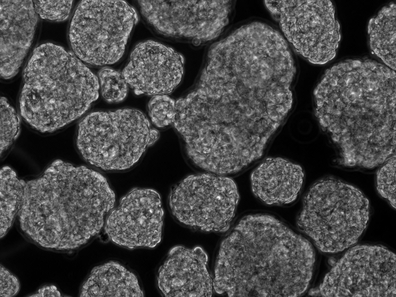 3D heart tissue constructs generated from stem cells. Credit: Antonio Rampoldi, Cardiomyocyte Stem Cell Laboratory, Emory University.
Technical Summary
Emory researchers generated 3D cardiac progenitors using Aggrewell 400 plates at differentiation day 6 and cryopreserved them at day 7. These samples were flown to the ISS through the SpaceX-20 mission. The astronauts on board the ISS thawed and cultured the cardiac progenitor spheres at 37°C with the final formulation of the CO2-independent medium. Live cultures were returned to the ground via warm storage after being cultured for 22 days on the ISS. The cardiac spheres had robust beating activity and majority of the cells in spheres were alive. These results demonstrate the feasibility of cryopreservation and CO2-independent culture of 3D cardiac progenitors for spaceflight experiments.
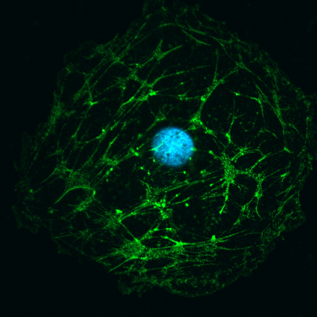 Stem cell–derived heart cell with staining of heart structure protein (green) and nucleus (blue). Credit: Antonio Rampoldi, Cardiomyocyte Stem Cell Laboratory, Emory University.
Developmental Stage
In vitro studies.
Read our featured innovation.
Patent Information
| App Type |
Country |
Serial No. |
Patent No. |
File Date |
Issued Date |
Patent Status |
| Utility (parent) |
United States |
17/344,654 |
|
6/10/2021 |
|
Pending |
|
|