Microfluidic Device Enhances Understanding of Clot Formation
Application
A microfluidic device for the measurement and quantification of the mechanical forces that occur during clot formation for use in hematology research or for diagnostic purposes.
Key Benefits
- Is capable of measuring platelet-platelet interactions at the single cell level that is not possible with current tools.
- Facilitates quick & easy analysis of clot formation for high throughput use.
- Is compatible with existing confocal microscopy technology and allows high-resolution imaging in real-time.
- Provides a platform for screening therapeutics that influence clot formation.
Market Summary
Microfluidic devices are a rapidly growing and important group of research tools. They are increasingly used in medical research, pharmaceutical research, and drug screening. These “lab-on-chip” type devices are built on microfluidics technology and integrate laboratory processes such as sample preparation, reactions, and analysis on a single miniature chip. Currently there are few microfluidic devices available for efficiently assessing the function of platelets and the mechanical forces at work during clot formation. Such a device would have applications not only as a research tool in the life sciences or drug development but also potentially as a diagnostic for blood or clotting disorders.
Technical Summary
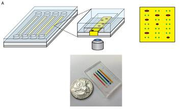
Abnormalities in clot formation are associated with thrombotic conditions such as cardiovascular disease and stroke. During clot formation, activated platelets interact with and attach to fibrin polymers. These interactions exert mechanical forces which can be measured and correspond to changes in the physical properties of the clot itself. Research in this field has been impeded by the inability to measure these platelet interactions on a cellular level as current research tools and methods are too large in scale. Emory investigators have created a three-layer microfluidic device that can measure platelet and fibrin interactions at the single-cell level. The bottom layer of the device consists of a hydrogel of a chosen stiffness. The next layer contains a collection of protein dots in a patterned alignment, and the third layer consists of microfluidic channels that can deliver a specific quantity of fluid or platelets to the layers below. Platelets delivered to the protein layer via the channels exert a contractile force on the protein dots and moves them out of their patterned alignment. These platelet interactions can be measured and the force exerted by the platelets can be calculated from this measurement. Because this process is contained in a single, standard-sized slide, the device is fully compatible with confocal microscopy which allows for high resolution and live-cell imaging. The high-throughput device is currently configured to measure 4 stiffnesses and 4 biochemical conditions, but may be scaled up to measure more conditions. With this tool, researchers can now gather a more comprehensive understanding of the mechanical properties of clot formation, identify potential therapeutic targets for cases in which platelet function is altered, or develop diagnostic tests based on platelet dynamics using the device.
Developmental Stage
Prototype device has been created and is currently used in laboratory research.
Patent Information
| App Type |
Country |
Serial No. |
Patent No. |
File Date |
Issued Date |
Patent Status |
| Utility (parent) |
United States |
14/202,946 |
10,168,341 |
3/10/2014 |
1/1/2019 |
Issued |
|
|